Cell Theory:
In 1833, German botanist Matthias Schleiden and German zoologist Theodor Schwann proposed that all plants and animals are composed of cells and that cells were the basic building blocks of life.
These observations led to the formulation of modern cell theory:
All organisms are made up of cells.
New cells are formed by the division of pre-existing cells.
Cells contains genetic material, which is passed on from parents to daughter cells.
All metabolic reactions take place inside the cells.
Exception to Cell Theory:
Viruses are puzzle in biology. Viruses, viroids and prions are the exception to cell theory. They lack protoplasm, the essential part of the cell and exists as obligate parasites which are sub-cellular in nature.
Types of cells:
On the basis of the cellular organization and the nuclear characteristics, the cell can be classified into Prokaryotes, Mesokaryotes and Eukaryotes.
Prokaryotes:
Those organisms with primitive nucleus are called as prokaryotes (pro – primitive; karyon – nucleus). The DNA lies in the ‘nucleoid’ which is not bound by the nuclear membrane and therefore it is not a true nucleus and is also a primitive type of nuclear material. The DNA is without histone proteins. Example: Bacteria, blue green algae, Mycoplasma, Rickettsiae and Spirochaetae.
Mesokaryotes:
In the year 1966, scientist Dodge and his coworkers proposed another kind of organisms called mesokaryotes. These organisms which shares some of the characters of both prokaryotes and eukaryotes. In other words these are organisms intermediate between pro and eukaryotes. These contains well organized nucleus with nuclear membrane and the DNA is organized into chromosomes but without histone protein components divides through amitosis similar with prokaryotes. Certain Protozoa like Noctiluca, some phytoplanktons like Gymnodinium, Peridinium and Dinoflagellates are representatives of mesokaryotes.
Eukaryotes:
Those organisms which have true nucleus are called Eukaryotes (Eu – True; karyon – nucleus). The DNA is associated with histones forming the chromosomes. Membrane bound organelles are present. Few organelles may have risen by endosymbiosis which is a cell living inside another cell. The Organelles like mitochondria and chloroplast well support this theory
Features of Prokaryotes, Mesokaryotes and Eukaryotes:
Features | Prokaryotes | Mesokaryotes | Eukaryotes |
Size of the cell | ~1-5µm | ~5-10µm | ~10-100µm |
Nuclear character | Nucleoid, no true nucleus, | Nucleus with nuclear membrane | True nucleus with nuclear membrane |
DNA | Usually circular without histone proteins | Usually linear but without histone proteins | Usually linear with histone proteins |
RNA/Protein synthesis | Couples in cytoplasm | Similar with eukaryotes | RNA synthesis inside nucleus/ Protein synthesis in cytoplasm |
Ribosomes | 50S+ 30S | 60S + 40S | 60S + 40S |
Organelles | Absent | Present | Numerous |
Cell movement | Flagella | Gliding and flagella | Flagella and cilia |
Organization | Usually single cell | Single and colony | Single, colonial and multicellular |
Cell division | Binary fission | Binary fission | Mitosis and meiosis |
Examples | Bacteria and Archaea | Dinoflagellate, Protozoa | Fungi, plants and animals |
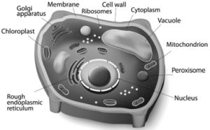
Plant and Animal cell:
An eukaryotic cell is highly distinct in its organisation. It shows several variations in different organisms. For instance, eukaryotic cells in plants and animals vary greatly.
Animal Cell:
Animal cells are surrounded by cell membrane or plasma membrane. Inside this membrane a gelatinous matrix called protoplasm is seen to contain nucleus and other organelles which include the endoplasmic reticulum, mitochondria, golgi bodies, centrioles, lysosomes, ribosomes and cytoskeleton.
Plant cell:
A typical plant cell has prominent cell wall, a large central vacuole and plastids in addition to other organelles present in animal cell.
Cell components and their functions:
S.No | Cell Components | Main Functions | Special Name |
1 | Cell wall | Surrounds and protects the cell Make the cell stiff and strong | Supporter and protector |
2 | Cell membrane | Holds and protects the cell Controls the movement of materials in and out of the cell | Gate of the cell |
3 | Cytoplasm | A watery, gel-like material in which cell parts move | Area of movement |
4 | Mitochondria | Produce and supply most of the energy for the cell | Power house of the cell |
5 | Chloroplasts | Contain green pigment chlorophyll Capture the energy of sunlight and use it to produce food for the cell by photosynthesis. | Food producers for the cell (Plant cell) |
6 | Vacuoles | Store food, water, and chemicals | Storage tanks |
7 | Nucleus | Acts as ‘brain’ of the cell Regulates and controls all the cell activities | Control centre |
8 | Nucleus membrane | Surrounds and protects the nucleus control the movement of materials in and out of the nucleus | Gate of the nucleus |
Difference between plant and animal cells:
S.No | Plant cell | Animal Cell |
1 | Usually they are larger than animal cells | Usually smaller than plant cells |
2 | Cell wall present in addition to plasma membrane and consists of middle lamellae, primary and secondary walls | Cell wall absent |
3 | Plasmodesmata present | Plasmodesmata absent |
4 | Chloroplast present | Chloroplast absent |
5 | Vacuole large and permanent | Vacuole small and temporary |
6 | Tonoplast present around vacuole | Tonoplast absent |
7 | Centrioles absent except motile cells of lower plants | Centrioles present |
8 | Nucleus present along the periphery of the cell | Nucleus at the centre of the cell |
9 | Lysosomes are rare | Lysosomes present |
10 | Storage material is starch grains | Storage material is a glycogen granules |
Protoplasm:
Protoplasm is the living content of cell that is surrounded by plasma membrane. It is a colourless material that exists throughout the cell together with cytoplasm, nucleus and other organelles. Protoplasm is composed of a mixture of small particles, such as ions, amino acids, monosaccharides, water, and macromolecules like nucleic acids, proteins, lipids and polysaccharides. It appears colourless, jelly like gelatinous, viscous elastic and granular. It appears foamy due to the presence of large number of vacuoles. It responds to the stimuli like heat, electric shock, chemicals and so on.
Cell Wall:
Cell wall is the outermost protective cover of the cell. It is present in bacteria, fungi and plants whereas it is absent in animal cell. It was first observed by Robert Hooke. It is an actively growing portion. It is made up of different complex material in various organism. In bacteria it is composed of peptidoglycan, in fungi chitin and fungal cellulose, in algae cellulose, galactans and mannans. In plants it is made up of cellulose, hemicellulose, pectin, lignin, cutin, suberin and silica. In plant, cell wall shows three distinct regions Primary wall, Secondary wall and Middle lamellae.
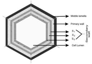
- Primary wall
It is the first layer inner to middle lamella, primarily consisting of loose network of cellulose microfibrils in a gel matrix. It is thin, elastic and extensible.In most plants the microfibrils are made up of cellulose oriented differently based on shape and thickness of the wall. The matrix of the primary wall is composed of hemicellulose, pectin, glycoprotein and water. Hemicellulose binds the microfibrils with matrix and glycoproteins control the orientation of microfibrils while pectin serves as filling material of the matrix. Cells such as parenchyma and meristems have only primary wall.
- Secondary wall
Secondary wall is laid during maturation of the cell. It plays a key role in determining the shape of a cell. It is thick, inelastic and is made up of cellulose and lignin. The secondary wall is divided into three sublayers termed as S1, S2 and S3 where the cellulose microfibrils are compactly arranged with different orientation forming a laminated structure and the cell wall strength is increased.
iii. Middle lamellae
It is the outermost layer made up of calcium and magnesium pectate, deposited at the time of cytokinesis. It is a thin amorphous layer which cements two adjacent cells. It is optically inactive (isotropic).
Plasmodesmata and Pits:
Plasmodesmata act as a channel between the protoplasm of adjacent cells through which many substances pass through. Moreover, at few regions, the secondary wall layer is laid unevenly whereas the primary wall and middle lamellae are laid continuously such regions are called pits. The Pits of adjacent cells are opposite to each other. Each pit has a pit chamber and a pit membrane. The pit membrane has many minute pores and thus they are permeable. The pits are of two types namely simple and bordered pit.
Functions of cell wall:
- Offers definite shape and rigidity to the cell.
- Serves as barrier for several molecules to enter the cells.
- Provides protection to the internal protoplasm against mechanical injury.
- Prevents the bursting of cells by maintaining the osmotic pressure.
- Plays a major role by acting as a mechanism of defense for the cells.
Cell Membrane:
The cell membrane is also called cell surface (or) plasma membrane. It is a thin structure which holds the cytoplasmic content called ‘cytosol’. It is extremely thin (less than 10nm).
Fluid Mosaic Model:
Jonathan Singer and Garth Nicolson (1972) proposed fluid mosaic model.
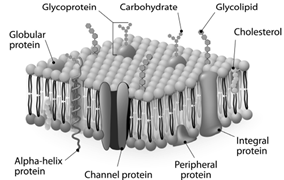
It is made up of lipids and proteins together with a little amount of carbohydrate. The lipid membrane is made up of phospholipid. The phospholipid molecule has a hydrophobic tail and hydrophilic head. The hydrophobic tail repels water and hydrophilic head attracts water. The proteins of the membrane are globular proteins which are found intermingled between the lipid bilayer most of which are projecting beyond the lipid bilayer. These proteins are called as integral proteins. Few are superficially attached on either surface of the lipid bilayer which are called as peripheral proteins. The proteins are involved in transport of molecules across the membranes and also act as enzymes, receptors (or) antigens.
Carbohydrate molecules of cell membrane are short chain polysaccharides. These are either bound with ‘glycoproteins’ or ‘glycolipids’ and form a ‘glyocalyx’.
The movement of membrane lipids from one side of the membrane to the other side by vertical movement is called flip flopping or flip flop movement. This movement takes place more slowly than lateral diffusion of lipid molecule. The Phospholipids can have flip flop movement because they have smaller Polar Regions, whereas the proteins cannot flip flop because the polar region is extensive.
Function of Cell Membrane:
The functions of the cell membrane is enormous which includes cell signalling, transporting nutrients and water, preventing unwanted substances entering into the cell, and so on.
Cytoplasm:
Cytoplasm is the main arena of various activities of a cell. It is the semifluid gelatinous substance that fills the cell. It is made up of eighty percent water and is usually clear and colourless. The cytoplasm is sometimes described as non nuclear content of protoplasm. The cytoplasm serves as a molecular soup where all the cellular organelles are suspended and bound together by a lipid bilayer plasma membrane. It constitutes dissolved nutrients, numerous salts and acids to dissolve waste products. It is a very good conductor of electricity. It gives support and protection to the cell organelles. It helps movement of the cellular materials around the cell through a process called cytoplasmic streaming. Further, most cellular activities such as many metabolic pathways including glycolysis and cell division occur in cytoplasm.
Cell Organelles:
Endomembrane System:
System of membranes in a eukaryotic cell, comprises the plasma membrane, nuclear membrane, endoplasmic reticulum, Golgi apparatus, lysosomes and vacuolar membranes (tonoplast). Endomembranes are made up of phospholipids with embedded proteins that are similar to cell membrane which occur within the cytoplasm. The endomembrane system is evolved from the inward growth of cell membrane in the ancestors of the first eukaryotes.
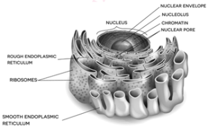
Endoplasmic Reticulum:
The largest of the internal membranes is called the endoplasmic reticulum (ER). The name endoplasmic reticulum was given by K.R. Porter (1948).
It consists of double membrane. Morphologically the structure of endoplasmic reticulum consists of the following:
- Cisternae are long, broad, flat, sac like structures arranged in parallel bundles or stacks to form lamella. The space between membranes of cisternae is filled with fluid.
- Vesicles are oval membrane bound vacuolar structure.
- Tubules are irregular in shape, branched, smooth walled, enclose a space
Endoplasmic reticulum is associated with nuclear membrane and cell surface membrane. It forms a network in cytoplasm and gives mechanical support to the cell. Its chemical environment enables protein folding and undergo modification necessary for their function. Misfolded proteins are pulled out and are degraded in endoplasmic reticulum. When ribosomes are present in the outer surface of the membrane it is called as rough endoplasmic reticulum (RER), when the ribosomes are absent in the endoplasmic reticulum it is called as smooth Endoplasmic reticulum (SER). Rough endoplasmic reticulum is involved in protein synthesis and smooth endoplasmic reticulum are the sites of lipid synthesis. The smooth endoplasmic reticulum contains enzymes that detoxify lipid soluble drugs, certain chemicals and other harmful compounds.
Golgi Body (Dictyosomes):
In 1898, Camillo Golgi visualized a netlike reticulum of fibrils near the nucleus, were named as Golgi bodies. In plant cells they are found as smaller vesicles termed as dictyosomes.
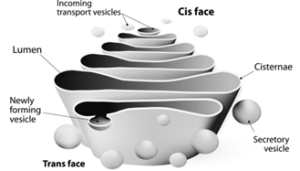
Golgi apparatus is a stack of flat membrane enclosed sacs. It consist of cisternae, tubules, vesicles and Golgi vacuoles. In plants, the cisternae are 10-20 in number placed in piles separated from each other by a thin layer of inter cisternal cytoplasm often flat or curved. Peripheral edge of cisternae forms a network of tubules and vesicles. Tubules interconnect cisternae and are 30-50nm in dimension. Vesicles are large round or concave sac. They are pinched off from the tubules.They are smooth/ secretary or coated type. Golgi vacuoles are large spherical structures filled with granular or amorphous substance, some function like lysosomes. Golgi apparatus compartmentalises a series of steps leading to the production of functional protein.
Small pieces of rough endoplasmic reticulum are pinched off at the ends to form small vesicles. A number of these vesicles then join up and fuse together to make a Golgi body. Golgi complex plays a major role in post translational modification of proteins and glycosylation of lipids.
Functions:
- Production of glycoproteins and glycolipids
- Transporting and storing of lipids.
- Formation of lysosomes.
- Production of digestive enzymes.
- Cell plate and cell wall formation
- Secretion of carbohydrates for the formation of plant cell walls and insect cuticles.
- Zymogen granules (proenzyme/precursor of all enzyme) are synthesised.
Mitochondria:
It was first observed by A. Kolliker (1880). Altmann (1894) named it as Bioplasts. Later Benda (1897, 1898), named as mitochondria. They are ovoid, rounded, rod shape and pleomorphic structures. Mitochondrion consists of double membrane, the outer and inner membrane. The outer membrane is smooth, highly permeable to small molecules and it contains proteins called Porins, which form channels that allows free diffusion of molecules smaller than about 1000 daltons and the inner membrane divides mitochondrion into two compartments, outer chamber between two membranes and the inner chamber is filled with matrix.
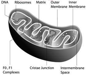
The inner membrane is convoluted (infoldings), called crista (plural: cristae). Cristae contain most of the enzymes for electron transport system. Inner chamber of the mitochondrion is filled with proteinaceous material called mitochondrial matrix. The Inner membrane consists of stalked particles called elementary particles or Fernandez Moran particles, F1 particles or Oxysomes. Each particle consists of a base, stem and a round head. In the head, ATP synthase is present for oxidative phosphorylation. Inner membrane is impermeable to most ions, small molecules and maintains the proton gradient that drives oxidative phosphorylation.
Mitochondria contain 73% of proteins, 25-30% of lipids, 5-7 % of RNA, DNA (in traces) and enzymes (about 60 types). Mitochondria are called Power house of a cell, as they produce energy rich ATP.
All the enzymes of Kreb’s cycle are found in the matrix except succinate dehydrogenase. Mitochondria consist of circular DNA and 70S ribosome. They multiply by fission and replicates by strand displacement model. Because of the presence of DNAs it is semiautonomous organelle. Unique characteristic of mitochondria is that they are inherited from female parent only. Mitochondrial DNA comparisons are used to trace human origins. It is also used to track and date recent evolutionary time because it mutates 5 to 10 time faster than DNA in the nucleus.
Chloroplast:
Chloroplasts are vital organelle found in green plants. Chloroplast has a double membrane the outer membrane and the inner membrane separated by a space called periplastidial space. The space enclosed by the inner membrane of chloroplast is filled with gelatinous matrix, lipo-proteinaceous fluid called stroma. Inside the stroma there are flat interconnected sacs called thylakoid. The membrane of thylakoid enclose a space called thylakoid lumen.
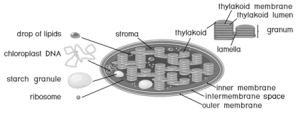
Grana (singular: Granum) are formed when many of these thylakoids are stacked together like pile of coins. Light is absorbed and converted into chemical energy in the granum, which is used in stroma to prepare carbohydrates. Thylakoid contain chlorophyll pigments. The chloroplast contains osmophilic granules, 70s ribosomes, DNA (circular and non histone) and RNA. These chloroplast genome encodes approximately 30 proteins involved in photosynthesis including the components of photosystem I & II, cytochrome bf complex and ATP synthase. One of the subunits of RuBisco is encoded by chloroplast DNA. It is the major protein component of chloroplast stroma, single most abundant protein on earth. The thylakoid contain small, rounded photosynthetic units called quantosomes. Chloroplast is a semi-autonomous organelle and divides by fission.
Functions:
- Photosynthesis
- Light reactions takes place in granum,
- Dark reactions take place in stroma,
- Chloroplast is involved in photo-respiration.
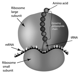
Ribosomes:
Ribosomes were first observed by George Palade (1953) as dense particles or granules in the electron microscope. Electron microscopic observation reveals that ribosomes are composed of two rounded sub units, united together to form a complete unit. Mg2+ is required for structural cohesion of ribosomes. Biogenesis of ribosome is a de nova formation, auto replication and nucleolar origin. Each ribosome is made up of one small and one large sub-unit Ribosomes are the sites of protein synthesis in the cell. Ribosome is not a membrane bound organelle.
Ribosome consists of RNA and protein: RNA 60 % and protein 40%. During protein synthesis, many ribosomes are attached to the single mRNA and is called polysomes or polyribosomes. The function of polysomes is the formation of several copies of a particular polypeptide during protein synthesis. They are free in non-protein synthesising cells. In protein synthesising cells they are linked together with the help of Mg2+ ions.
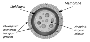
Lysosomes (Suicidal Bags of Cell):
Lysosomes were discovered by Christian de Duve (1953), these are known as suicidal bags. They are spherical bodies enclosed by a single unit membrane. They are found in eukaryotic cell. Lysosomes are small vacuoles formed when small pieces of golgi body are pinched off from its tubules.
They contain a variety of hydrolytic enzymes that can digest material within the cell. The membrane around lysosome prevent these enzymes from digesting the cell itself.
Functions:
- Intracellular digestion: They digest carbohydrates, proteins and lipids present in cytoplasm.
- Autophagy: During adverse condition they digest their own cell organelles like mitochondria and endoplasmic reticulum
- Autolysis: Lysosome causes self destruction of cell.
- Ageing: Lysosomes have autolytic enzymes that disrupts intracellular molecules.
- Phagocytosis: Large cells or contents are engulfed and digested by macrophages, thus forming a phagosome in cytoplasm. These phagosome fuse with lysosome for further digestion.
- Exocytosis: Lysosomes release their enzymes outside the cell to digest other cells.
Microbodies:
Eukaryotic cells contain many enzyme bearing membrane enclosed vesicles called microbodies. They are single unit membrane bound cell organelles. Example: Peroxisomes and glyoxysomes.
Peroxisomes:
Peroxisomes were identified as organelles by Christian de Duve (1967). Peroxisomes are small spherical bodies and single membrane bound organelle. It takes part in photorespiration and associated with glycolate metabolism. In plants, leaf cells have many peroxisomes. It is also commonly found in liver and kidney of mammals. These are also found in cells of protozoa and yeast.
Glyoxysomes:
Glyoxysome was discovered by Harry Beevers (1961). It is a single membrane bound organelle. It is a sub cellular organelle and contains enzymes of glyoxylate pathway. β-oxidation of fatty acid occurs in glyoxysomes of germinating seeds Example: Castor seeds.
Sphaerosomes:
It is spherical in shape and enclosed by single unit membrane. Example: Storage of fat in the endosperm cells of oil seeds.
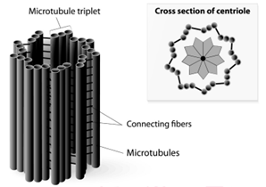
Centrioles:
Centrioles consists of nine triplet peripheral fibrils made up of tubulin. The central part of the centriole is called hub, is connected to the tubules of the peripheral triplets by radial spokes (9+0 pattern). The centriole form the basal body of cilia or flagella and spindle fibers which forms the spindle apparatus in animal cells. The membrane is absent in centriole (non-membranous organelle).
Vacuoles:
In plant cells vacuoles are large, bounded by a single unit membrane called Tonoplast. The Vacuoles contain cell sap, which is a solution of sugars, amino acids, mineral salts, waste chemical and anthocyanin pigments. Beetroot cells contain anthocyanin pigments in their vacuoles. Vacuoles accumulate products like tannins. The osmotic expansion of a cell kept in water is chiefly regulated by vacuole and the water enters the vacuole by osmosis.
The major function of plant vacuole is to maintain water pressure known as turgor pressure, which maintains the plant structure. Vacuoles organises itself into a storage/ sequestration compartment. Example: Vacuoles store, most of the sucrose of the cell.
Example: Sugar in Sugar beet and Sugar cane.
Nucleus:
Nucleus is an important unit of cell which controls all activities of the cell. Nucleus holds the hereditary information. It is the largest among all cell organelles. It may be spherical, cuboidal, ellipsoidal or discoidal.
It is surrounded by a double membrane structure called nuclear envelope, which has the inner and outer membrane. The inner membrane is smooth without ribosomes and the outer membrane is rough by the presence of ribosomes and it continues with irregular and infrequent intervals with the endoplasmic reticulum. The membrane is perforated by pores known as nuclear pores which allows materials such as mRNA, ribosomal units, proteins and other macromolecules to pass in and out of the nucleus. The pores enclosed by circular structures called annuli. The pore and annuli form the pore complex. The space between two membranes is called perinuclear space.
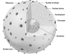
Nuclear space is filled with nucleoplasm, a gelatinous matrix has uncondensed chromatin network and a conspicuous nucleolius. The Chromatin network is an uncoiled, indistinct and remain thread like during the interphase. It has little amount of RNA and DNA bound to histone proteins in eukaryotic cells.
During cell division chromatin is condensed into an organized form called chromosome. The portion an eukaryotic chromosome which is transcribed into mRNA contains active genes that are nottightly condensed during interphase is called Euchromatin. The portion of an eukaryotic chromosome that is not transcribed into mRNA which remains condensed during interphase and stains intensely is called Heterochromatin. Nucleolus is a small, dense, spherical structure either present singly or in multiples inside the nucleus and it’s not membrane bound. Nucleoli possess genes for rRNA and tRNA.
Functions of the nucleus:
- Controlling all cellular activities
- Storing the genetic or hereditary information.
- Coding the information from DNA for the production of enzymes and proteins.
- DNA duplication and transcription takes place in the nucleus.
- In nucleolus ribosomal biogenesis takes place.
Chromosomes:
Strasburger (1875) first reported its present in eukaryotic cell and the term ‘chromosome’ was introduced by Waldeyer in 1888. Bridges (1916) first proved that chromosomes are the physical carriers of genes. It is made up of DNA and associated proteins.
Structure of chromosome:
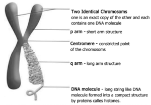
The chromosomes are composed of thread like strands called chromatin which is made up of DNA, protein and RNA. Each chromosome consists of two symmetrical structures called chromatids. During cell division the chromatids forms a well organized chromosomes with definite size and shape. They are identical and are called sister chromatids. A typical chromosome has narrow zones called constrictions. There are two types of constrictions, namely primary constriction and secondary constriction. The primary constriction is made up of centromere and kinetochore. Both the chromatids are united at centromere, whose number varies. The monocentric chromosome has one centromere and the polycentric chromosome has many centromeres. Centromere contains a complex system of protein fibres called kinetochore. Kinetochore is the region of chromosome which is attached to the spindle fibre during mitosis.
Besides primary there are few secondary constrictions, are present. Nucleoli develop from these secondary constrictions are called nucleolar organizers. Secondary constrictions contain the genes for ribosomal RNA which induce the formation of nucleoli and are called nucleolar organizer regions.
A satellite or SAT Chromosome is a short chromosomal segment or rounded body separated from main chromosome by a relatively elongated secondary constriction. It is a morphological entity in certain chromosomes.
Telomere is the terminal part of chromosome. It offers stability to the chromosome. DNA of the telomere has specific sequence of nucleotides. Telomere in all eukaryotes are composed of many repeats of short DNA sequences (5’TTAGGG3’ sequence in Neurospora crassa and human beings). Maintenance of telomeres appears to be an important factor in determining the life span and reproductive capacity of cells, so studies of telomeres and telomerase have the promise of providing new insights into conditions such as ageing and cancer. Telomeres prevent the fusion of chromosomal ends with one another.
Types of Chromosomes:
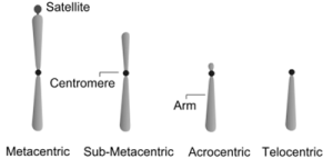
Based on the position of centromere, chromosomes are called telocentric (terminal centromere), acrocentric (terminal centromere capped by telomere), sub metacentric (centromere subterminal) and metacentric (centromere median). The eukaryotic chromosome may be rod shaped (telocentric and acrocentric), L-shaped (sub-metacentric) and V-shaped (metacentric).
Based on the functions of chromosome it can be divided into autosomes and sex chromosomes.Autosomes are present in all cells controlling somatic characteristics of an organism. In human diploid cell, 44 chromosomes are autosomes whereas two are sex chromosomes. Sex chromosomes are involved in the determination of sex.
Special types of chromosomes:
These chromosomes are larger in size and are called giant chromosomes in certain plants and they are found in the suspensors of the embryo. The polytene chromosome and lamp brush chromosome occur in animals and are also called as giant chromosomes.
Polytene chromosomes:
Polytene chromosomes observed in the salivary glands of Drosophila (fruit fly) by E.G. Balbiani in 1881. In larvae of many flies, midges (Dipthera) and some insects the interphase chromosomes duplicates and reduplicates without nuclear division. A single chromosome which is present in multiple copies form a structure called polytene chromosome which can be seen in light microscope.
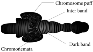
They are genetically active. There is a distinct alternating dark bands and light inter-bands. About 95% of DNA are present in bands and 5% in inter-bands. The polytene chromosome has extremely large puff called Balbiani rings which is seen in Chironomous larvae. It is also known as chromosomal puff. Puffing of bands are the sites of intense RNA synthesis. As this chromosome occurs in the salivary gland it is known as salivary gland chromosomes. Gene expression, transcription of genes and RNA synthesis occurs in the bands along the polytene chromosomes.
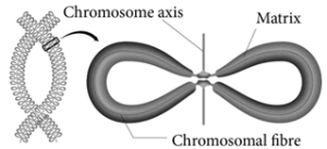
Lampbrush chromosomes:
Lampbrush chromosomes occur at the diplotene stage of first meiotic prophase in oocytes of an animal Salamandar and in giant nucleus of the unicellular alga Acetabularia. It was first observed by Flemming in 1882. The highly condensed chromosome forms the chromosomal axis, from which lateral loops of DNA extend as a result of intense RNA synthesis.
Cell Cycle:
Definition: A series of events leading to the formation of new cell is known as cell cycle. The series of events include several phases.
Duration of Cell Cycle:
Different kinds of cells have varied duration for cell cycle phases. Eukaryotic cell divides every 24 hours. The cell cycle is divided into mitosis and interphase. In a cell cycle 95% is spent for interphase whereas the mitosis and cytokinesis last only for an hour.
Cell cycle of a proliferating human cell | |
Phase | Time duration (in hrs) |
G1 | 11 |
S | 8 |
G2 | 4 |
M | 1 |
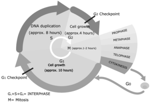
Interphase:
Longest part of the cell cycle, but it is of extremely variable length. At first glance the nucleus appears to be resting but this is not the case at all. The chromosomes previously visible as thread like structure, have dispersed. Now they are actively involved in protein synthesis, at least for most of the interphase.
G1 Phase:
The first gap phase – 2C amount of DNA in cells of G1. Cells become metabolically active and grows by producing proteins, lipids, carbohydrates and cell organelles including mitochondria and endoplasmic reticulum. Many checkpoints control the cell cycle. The check point are also called as the restriction point. First check point at the end of G1, determines a cells fate whether it will continue in the cell cycle and divide or enter a stage called G0 a quiescent stage, probably as specified cell or die. Cells are arrested in G1 due to:
- Nutrient deprivation
- Lack of growth factors or density dependant inhibition
- Undergo metabolic changes and enter into G0 state.
Biochemicals inside cell activates the cell division. The proteins called kinases and cyclins activate genes and their proteins to perform cell division. Cyclins act as major checkpoint which operates in G1 to determine whether or not a cell divides.
G0 Phase:
Some cells exit G1 and enters a quiescent stage called G0, where the cell remains metabolically active without proliferation. Cells can exist for long periods in G0 phase. In G0, cells cease growth with reduced rate of RNA and protein synthesis. The G0 phase is not permanent. Mature neuron and skeletal muscle cell remain permanently in G0. Many cells in animals remains in G0 unless called on to proliferate by appropriate growth factors or other extracellular signals. G0 cells are not dormant.
S phase – Synthesis phase – cells with intermediate amounts of DNA
Growth of the cell continues as replication of DNA occur, protein molecules called histones are synthesised and attach to the DNA. The centrioles duplicate in the cytoplasm. DNA content increases from 2C to 4C.
G2 – The second Gap phase – 4C amount of DNA in cells of G2 and mitosis
Cell growth continues by protein and cell organelle synthesis, mitochondria and chloroplasts divide. DNA content remains as 4C. Tubulin is synthesised and microtubules are formed. Microtubles organise to form spindle fibre. The spindle begins to form and nuclear division follows.
One of the proteins synthesized only in the G2 period is known as Maturation Promoting Factor (MPF). It brings about condensation of interphase chromosomes into the mitotic form. DNA damage checkpoints operates in G1 S and G2 phases of the cell cycle.
Cell Division:
Amitosis (Direct Cell Division):
Amitosis is also called direct or incipient cell division. Here there is no spindle formation and chromatin material does not condense. It consist of two steps.
Karyokinesis:
- Involves division of nucleus.
- Nucleus develops a constriction at the center and becomes dumbell shaped.
- Constriction deepens and divides the nucleus into two.
Cytokinesis:
- Involves division of cytoplasm.
- Plasma membrane develops a constriction along nuclear constriction.
- It deepens centripetally and finally divides the cell into two cells.
Example:
Cells of mammalian cartilage, macronucleus of Paramecium and old degenerating cells of higher plants.
Drawbacks of Amitosis:
- Causes unequal distribution of chromosomes.
- Can lead to abnormalities in metabolism and reproduction.
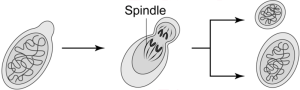
Mitosis:
Mitosis occurs in shoot and root tips and other meristematic tissues of plants associated with growth. The number of chromosomes in the parent and the daughter (Progeny) cells remain the same so it is also called as equational division.
Closed Mitosis:
In closed mitosis, the nuclear envelope remains intact and chromosomes migrate to opposite poles of a spindle within the nucleus. Example: Many single celled eukaryotes including yeast and slime molds.
Open Mitosis:
In open mitosis, the nuclear envelope breaks down and then reforms around the 2 sets of separated chromosome. Example: Most plants and animals.
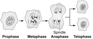
Stages of Mitosis:
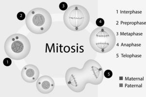
- Prophase
Prophase is the longest phase in mitosis. Chromosomes become visible as long thin thread like structure, condenses to form compact mitotic chromosomes. In plant cells initiation of spindle fibres takes place, nucleolus disappears. Nuclear envelope breaks down. Golgi apparatus and endoplasmic reticulum disappear.
In animal cell the centrioles extend a radial array of microtubules and reach the poles of the cell. This arrangement of microtubules is called an aster. Plant cells do not form asters.
- Metaphase
Chromosomes (two sister chromatids) are attached to the spindle fibres by kinetochore of the centromere. The spindle fibres are made up of tubulin. The alignment of chromosome into compact group at the equator of the cell is known as metaphase plate. This is the stage where the chromosomal morphology can be easily studied.
Kinetochore is a DNA–Protein complex present at the centromere where microtubules are attached. It is a trilaminar disc like plate.
iii. Anaphase
Each chromosome splits simultaneously and two daughter chromatids begin to migrate towards two opposite poles of a cell. Each centromere splits longitudinally into two, freeing the two sister chromatids from each other. When sister chromatids separate the actual partitioning of the replicated genome is complete.
APC (Anaphase Promoting Complex) is a cluster of proteins that induces the breaking down of cohesion proteins which leads to the separation of chromatids during mitosis. Thus it helps in the transition of metaphase to anaphase.
- Telophase
Two sets of daughter chromosomes reach opposite poles of the cell and mitotic spindle disappears. Division of genetic material is completed during karyokinesis, followed by cytokinesis (division of cytoplasm). Nucleolus and nuclear membranes reforms. Nuclear membrane form around each set of chromosomes. Now the chromosomes decondense. In plants, phragmoplast are formed between the daughter cells. Cell plate is formed between the two daughter cells, reconstruction of cell wall takes place. Finally cells are separated by the distribution of organelles, macromolecules into two newly formed daughter cells.
Cytokinesis:
Cytokinesis in Animal Cells:
It is a contractile process. The ring consists of a bundle of microfilaments assembled from actin and myosin. This fibril generates a contractile force, that draws the ring inward forming a cleavage furrow in the cell. Thus it divides the cell into two.
Cytokinesis in Plant Cell:
Division of the cytoplasm often starts during telophase. In plants, cell plate grows from center towards lateral walls.
Phragmoplast contains microtubules, actin filaments and vesicles from golgi apparatus and ER. Microtubule of the pharagmoplast move to the equator, fuse to form a new plasma membrane and the materials which are placed there becomes new cell wall. The first stage of cell wall construction is a line dividing the newly forming cells called a cell plate. The cell plate eventually stretches right across the cell forming the middle lamella. Cellulose builds up on each side of the middle lamella to form the cell walls of two new plant cells.
Meiosis:
In Greek meioum means to reduce. Meiosis is unique because of synapsis, homologous recombination and reduction division. Meiosis takes place in the reproductive organs. It results in the formation of gametes with half the normal chromosome number.
Haploid sperms are made in testes; haploid eggs are made in ovaries of animals.
In flowering plants meiosis occurs during microsporogenesis in anthers and megasporogenesis in ovule. In contrast to mitosis, meiosis produces cells that are not genetically identical. So meiosis has a key role in producing new genetic types which results in genetic variation.
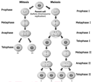
Stages in Meiosis:
Meiosis can be studied under two divisions i.e., meiosis I and meiosis II. As with mitosis, the cell is said to be in interphase when it is not dividing.
Meiosis I-Reduction Division:
Prophase I – Prophase I is of longer duration and it is divided into 5 substages – Leptotene, Zygotene, Pachytene, Diplotene and Diakinesis.
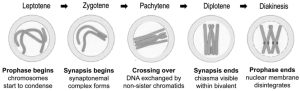
- Leptotene – Chromosomes are visible under light microscope. Condensation of chromosomes takes place. Paired sister chromatids begin to condense.
- Zygotene – Pairing of homologous chromosomes takes place and it is known as synapsis. Chromosome synapsis is made by the formation of synaptonemal complex. The complex formed by the homologous chromosomes are called as bivalent (tetrads).
iii. Pachytene – At this stage bivalent chromosomes are clearly visible as tetrads. Bivalent of meiosis I consists of 4 chromatids and 2 centromeres. Synapsis is completed and recombination nodules appear at a site where crossing over takes place between non-sister chromatids of homologous chromosome. Recombination of homologous chromosomes is completed by the end of the stage but the chromosomes are linked at the sites of crossing over. This is mediated by the enzyme recombinase.
- Diplotene – Synaptonemal complex disassembled and dissolves. The homologous chromosomes remain attached at one or more points where crossing over has taken place. These points of attachment where ‘X’ shaped structures occur at the sites of crossing over is called Chiasmata. Chiasmata are chromatin structures at sites where recombination has been taken place. They are specialised chromosomal structures that hold the homologous chromosomes together. Sister chromatids remain closely associated whereas the homologous chromosomes tend to separate from each other but are held together by chiasmata. This substage may last for days or years depending on the sex and organism.
- Diakinesis – Terminalisation of chiasmata, homologous chromosomes become short and condensed. Nucleolus and nuclear envelope disappears. Spindle fibres assemble.
Metaphase I:
Spindle fibres are attached to the centromeres of the two homologous chromosomes. Bivalent (pairs of homologous chromosomes) aligned at the equator of the cell known as metaphase plate.
The random distribution of homologous chromosomes in a cell in Metaphase I is called independent assortment.
Anaphase I:
Homologous chromosomes are separated from each other by shortening of spindle fibers. Each homologous chromosomes with its two chromatids and undivided centromere move towards the opposite poles of the cells. The actual reduction in the number of chromosomes takes place at this stage. Homologous chromosomes which move to the opposite poles are either paternal or maternal in origin. Sister chromatids remain attached with their centromeres.
Telophase I:
Haploid set of chromosomes are present at each pole. The formation of two daughter cells, each with haploid number of chromosomes takes place. Nuclei reassembled. Nuclear envelope forms around the chromosome and the chromosomes becomes uncoiled. Nucleolus reappears.
In plants after karyokinesis, cytokinesis takes place by which two daughter cells are formed by the cell plate between 2 groups of chromosomes known as dyad of cells (haploid).
The stage between the two meiotic divisions is called interkinesis which is short-lived.
Meiosis II – Equational division:
This division is otherwise called mitotic meiosis because it resembles mitosis. Since it includes all the stages of mitotic divisions.
Prophase II:
The chromosome with 2 chromatids becomes short, condensed, thick and becomes visible. New spindle develops at right angles to the cell axis. Nuclear membrane and nucleolus disappear.
Metaphase II:
Chromosome arranged at the equatorial plane of the spindle. Microtubules of spindle gets attached to the centromere of sister chromatids.
Anaphase II:
Sister chromatids separate. The daughter chromosomes move to the opposite poles due to shortening of spindle fibres. Centromere of each chromosome split, allowing to move towards opposite poles of the cells holding the sister chromatids.
Telophase II:
Four groups of chromosomes are organised into four haploid nuclei. The spindle disappears. Nuclear envelope, nucleolus reappear.
After karyokinesis, cytokinesis follows and four haploid daughter cells are formed, called tetrads.
Significance of Meiosis:
This maintains a definite constant number of chromosomes in organisms.
Crossing over takes place and exchange of genetic material leads to variations among species. These variations are the raw materials to evolution. Meiosis leads to genetic variability by partitioning different combinations of genes into gametes through independent assortment.
Adaptation of organisms to various environmental stress.
Semi-autonomous organelles:
Semi-autonomous organelles are those organelles that contain their own DNA and reproduce independently of the nucleus. Chloroplast and mitochondria are called semi-autonomous organelles because they have their own genetic material (DNA) and are capable of synthesizing proteins required for their functioning.