Functions, mode of absorption and deficiency symptoms of macronutrients:
Macronutrients, their functions, their mode of absorption, deficiency symptoms and deficiency diseases are discussed here:
Nitrogen (N): It is required by the plants in greatest amount. It is an essential component of proteins, nucleic acids, amino acids, vitamins, hormones, alkaloids, chlorophyll and cytochrome. It is absorbed by the plants as nitrates (NO3).
Deficiency symptoms: Chlorosis, stunted growth, anthocyanin formation.
Phosphorus (P): Constituent of cell membrane, proteins, nucleic acids, ATP, NADP, phytin and sugar phosphate. It is absorbed as H2PO41 and HPO42 ions.
Deficiency symptoms: Stunted growth, anthocyanin formation, and necrosis, inhibition of cambial activity, affect root growth and fruit ripening.
Potassium (K): Maintains turgidity and osmotic potential of the cell, opening and closure of stomata, phloem translocation, stimulate activity of enzymes, anion and cation balance by ion-exchange. It is absorbed as K1 ions.
Deficiency symptoms: Marginal chlorosis, necrosis, low cambial activity, loss of apical dominance, lodging in cereals and curled leaf margin.
Calcium (Ca): It is involved in synthesis of calcium pectate in middle lamella, mitotic spindle formation, mitotic cell division, and permeability of cell membrane, lipid metabolism, activation of phospholipase, ATPase, amylase and activator of adenyl kinase. It is absorbed as Ca21 exchangeable ions.
Deficiency symptoms: Chlorosis, necrosis, stunted growth, premature fall of leaves and flowers, inhibit seed formation, Black heart of Celery, Hooked leaf tip in Sugar beet, Musa and Tomato.
Magnesium (Mg): It is a constituent of chlorophyll, activator of enzymes of carbohydrate metabolism (RUBP Carboxylase and PEP Carboxylase) and involved in the synthesis of DNA and RNA. It is essential for binding of ribosomal sub units. It is absorbed as Mg21 ions.
Deficiency symptoms: Inter veinal chlorosis, necrosis, anthocyanin (purple) formation and Sand drown of tobacco.
Sulphur (S): Essential component of amino acids like cystine, cysteine and methionine, constituent of coenzyme A, Vitamins like biotin and thiamine, constituent of proteins and ferredoxin. Plants utilise sulphur as sulphate (SO42) ions.
Deficiency symptoms: Chlorosis, anthocyanin formation, stunted growth, rolling of leaf tip and reduced nodulation in legumes.
Functions, mode of absorption and deficiency symptoms of micronutrients:
Micronutrients even though required in trace amounts are essential for the metabolism of plants. They play key roles in many plants. Example: Boron is essential for translocation of sugars, molybdenum is involved in nitrogen metabolism and zinc is needed for biosynthesis of auxin. Here, we will study about the role of micro nutrients, their functions, their mode of absorption, deficiency symptoms and deficiency diseases.
Iron (Fe): Iron is required lesser than macronutrient and larger than micronutrients, hence, it can be placed in any one of the groups. Iron is an essential element for the synthesis of chlorophyll and carotenoids. It is the component of cytochrome, ferredoxin, flavoprotein, formation of chlorophyll, porphyrin, activation of catalase, peroxidase enzymes. It is absorbed as ferrous (Fe21) and ferric (Fe31) ions. Absorption of Fe2+ ions are comparatively more than Fe3+ ions. Mostly fruit trees are sensitive to iron.
Deficiency: Interveinal Chlorosis, formation of short and slender stalk and inhibition of chlorophyll formation.
Manganese (Mn): Activator of carboxylases, oxidases, dehydrogenases and kinases, involved in splitting of water to liberate oxygen (photolysis). It is absorbed as manganous (Mn21) ions.
Deficiency: Interveinal chlorosis, grey spot on oats leaves and poor root system.
Copper(Cu): Constituent of plastocyanin, component of phenolases, tyrosinase, enzymes involved in redox reactions, synthesis of ascorbic acid, maintains carbohydrate and nitrogen balance, part of oxidase and cytochrome oxidase. It is absorbed as cupric (Cu21) ions.
Deficiency: Die back of citrus, Reclamation disease of cereals and legumes, chlorosis, necrosis and Exanthema in Citrus.
Zinc (Zn): Essential for the synthesis of Indole acetic acid (Auxin), activator of carboxylases, alcohol dehydrogenase, lactic dehydrogenase, glutamic acid dehydrogenase, carboxy peptidases and tryptophan synthetase. It is absorbed as Zn21 ions.
Deficiency: Little leaf and mottle leaf due to deficiency of auxin, Inter veinal chlorosis, stunted growth, necrosis and Khaira disease of rice.
Boron(B): Translocation of carbohydrates, uptake and utilisation of Ca11, pollen germination, nitrogen metabolism, fat metabolism, cell elongation and differentiation. It is absorbed as (borate) BO32 ions.
Deficiency: Death of root and shoot tips, premature fall of flowers and fruits, brown heart of beet root, internal cork of apple and fruit cracks.
Molybdenum (Mo): Component of nitrogenase, nitrate reductase, involved in nitrogen metabolism, and nitrogen fixation. It is absorbed as molybdate (Mo21) ions.
Deficiency: Chlorosis, necrosis, delayed flowering, retarded growth and whip tail disease of cauliflower.
Chlorine (Cl): It is involved in Anion – Cation balance, cell division, photolysis of water. It is absorbed as Cl2 ions.
Deficiency: Wilting of leaf tips
Nickel (Ni): Cofactor for enzyme urease and hydrogenase.
Deficiency: Necrosis of leaf tips.
Special modes of nutrition:
Nutrition is the process of uptake and utilization of nutrients by living organisms. There are two main types such as autotrophic and heterotrophic nutrition. Autotrophic nutrition is further divided into photosynthetic and chemosynthetic nutrition. Heterotrophic nutrition is further divided into saprophytic, parasitic, symbiotic and insectivorous type. In this topic you are going to learn about special mode of nutrition.
Saprophytic mode of nutrition in angiosperms:
Saprophytes derive nutrients from dead and decaying matter. Bacteria and fungus are main saprophytic organisms. Some angiosperms also follow saprophytic mode of nutrition. Example: Neottia. Roots of Neottia (Bird’s Nest Orchid) associate with mycorrhizae and absorb nutrients as a saprophyte. Monotropa (Indian Pipe) grow on humus rich soil found in thick forests. It absorbs nutrient through mycorrhizal association.
Parasitic mode of nutrition in angiosperms:
Organisms deriving their nutrient from another organism (host) and causing disease to the host are called parasites.
Obligate or Total parasite – Completely depends on host for their survival and produces haustoria.
Total stem parasite: The leafless stem twine around the host and produce haustoria. Example: Cuscuta (Dodder), a rootless plant growing on Zizyphus, Citrus and so on.
Total root parasite: They do not have stem axis and grow in the roots of host plants produce haustoria. Example: Rafflesia, Orobanche and Balanophora.
Partial parasite – Plants of this group contain chlorophyll and synthesize carbohydrates. Water and mineral requirements are dependent on host plant.
Partial Stem Parasite: Example: Loranthus and Viscum (Mistletoe)
Loranthus grows on fig and mango trees and absorb water and minerals from xylem.
Partial root parasite: Example: Santalum album (Sandal wood tree) in its juvenile stage produces haustoria which grows on roots of many plants (Figure 12.10).
3 Symbiotic mode of Nutrition:
Lichens: It is a mutual association of Algae and Fungi. Algae prepares food and fungi absorbs water and provides thallus structure.
Mycorrhizae: Fungi associated with roots of higher plants including Gymnosperms. Example: Pinus.
Rhizobium and Legumes: This symbiotic association fixes atmospheric nitrogen
Cyanobacteria and Coralloid Roots: This association is found in Cycas where Nostoc associates with its coralloid roots.
Insectivorous mode of nutrition:
Plants which are growing in nitrogen deficient areas develop insectivorous habit to resolve nitrogen deficiency. These plants obtain nitrogen from the insects
Nepenthes (Pitcher plant): Pitcher is a modified leaf and contains digestive enzymes. Rim of the pitcher is provided with nectar glands and acts as an attractive lid. When insect is trapped, proteolytic enzymes will digest the insect.
Drosera (Sundew): It consists of long club shaped leaves with tentacles that secrete sticky digestive fluid which looks like a sundew and attracts insects.
Utricularia (Bladderwort): Submerged plant in which leaf is modified into a bladder to collect insect in water.
Dionaea (Venus fly trap): Leaf of this plant modified into a colourful trap. Two folds of lamina consist of sensitive trigger hairs and when insects touch the hairs it will close and traps the insects.
Nutritional and digestive disorders:
Intestinal tract is more prone to bacterial, viral and parasitic worm infections. This infection may cause inflammation of the inner lining of colon called colitis. The most common symptoms of colitis are rectal bleeding, abdominal cramps, and diarrhoea.
Protein energy malnutrition: (PEM)
Growing children require more amount of protein for their growth and development. Protein deficient diet during early stage of children may lead to protein energy malnutrition such as Marasmus and Kwashiorkor. Symptoms are dry skin, pot-belly, oedema in the legs and face, stunted growth, changes in hair colour, weakness and irritability. Marasmus is an acute form of protein malnutrition. This condition is due to a diet with inadequate carbohydrate and protein. Such children are suffer from diarrhoea, body becomes lean and weak (emaciated) with reduced fat and muscle tissue with thin and folded skin.
Indigestion: It is a digestive disorder in which the food is not properly digested leading to a feeling of fullness of stomach. It may be due to inadequate enzyme secretion, anxiety, food poisoning, over eating, and spicy food.
Constipation: In this condition, the faeces are retained within the rectum because of irregular bowel movement due to poor intake of fibre in the diet and lack of physical activities.
Vomiting: It is reverse peristalsis. Harmful substances and contaminated food from stomach are ejected through the mouth. This action is controlled by the vomit centre located in the medulla oblongata. A feeling of nausea precedes vomiting.
Jaundice: It is the condition in which liver is affected and the defective liver fails to break down haemoglobin and to remove bile pigments from the blood. Deposition of these pigments changes the colour of eye and skin yellow. Sometimes, jaundice is caused due to hepatitis viral infections.
Liver cirrhosis: Chronic disease of liver results in degeneration and destruction of liver cells resulting in abnormal blood vessel and bile duct leading to the formation of fibrosis. It is also called deserted liver or scarred liver. It is caused due to infection, consumption of poison, malnutrition and alcoholism.
Gall Stones: Any alteration in the composition of the bile can cause the formation of stones in the gall bladder. The stones are mostly formed of crystallized cholesterol in the bile. The gall stone causes obstruction in the cystic duct, hepatic duct and also hepato-pancreatic duct causing pain, jaundice and pancreatitis.
Appendicitis: It is the inflammation of the vermiform appendix, leading to severe abdominal pain. The treatment involves the removal of appendix by surgery. If treatment is delayed the appendix may rupture and results in infection of the abdomen, called peritonitis.
Hiatus hernia (Diaphragmatic hernia): It is a structural abnormality in which superior part of the stomach protrudes slightly above the diaphragm. The exact cause of hiatus hernias is not known. In some people, injury or other damage may weaken muscle tissue, by applying too much pressure (repeatedly) on the muscles around the stomach while coughing, vomiting, and straining during bowel movement and lifting heavy object. Heart burn is also common in those with a hiatus hernia. In this condition, stomach contents travel back into the oesophagus or even into oral cavity and causes pain in the centre of the chest due to the eroding nature of acidity.
Diarrhoea: It is the most common gastrointestinal disorder worldwide. It is sometimes caused by bacteria or viral infections through food or water. When the colon is infected, the lining of the intestine is damaged by the pathogens, thereby the colon is unable to absorb fluid. The abnormal frequency of bowel movement and increased liquidity of the faecal discharge is known as diarrhoea. Unless the condition is treated, dehydration can occur. Treatment is known as oral hydration therapy. This involves drinking plenty of fluids – sipping small amounts of water at a time to rehydrate the body.
Peptic ulcer: It refers to an eroded area of the tissue lining (mucosa) in the stomach or duodenum. Duodenal ulcer occurs in people in the age group of 25 – 45 years. Gastric ulcer is more common in persons above the age of 50 years. Ulcer is mostly due to infections caused by the bacterium Helicobacter pylori. It may also be caused due to uncontrolled usage of aspirin or certain anti-inflammatory drugs. Ulcer may also be caused due to smoking, alcohol, caffeine and psychological stress.
Obesity: It is caused due to the storage of excess of body fat in adipose tissue. It may induce hypertension, atherosclerotic heart disease and diabetes. Obesity may be genetic or due to excess intake of food, endocrine and metabolic disorders. Degree of obesity is assessed by body mass index (BMI). A normal BMI range for adult is 19-25 above 25 is considered as obese. BMI is calculated as body weight in Kg, divided by the square of body height in meters. For example, a 50 Kg person with a height of 160 cms would have a BMI of 19.5.
That is BMI = 50 / (1.6)2 = 19.5
Disorders related to the Excretory System:
Urinary tract infection:
Female’s urethra is very short and its external opening is close to the anal opening, hence improper toilet habits can easily carry faecal bacteria into the urethra. The urethral mucosa is continuous with the urinary tract and the inflammation of the urethra (urethritis) can ascend the tract to cause bladder inflammation (cystitis) or even renal inflammation (pyelitis or pyelonephritis). Symptoms include dysuria (painful urination), urinary urgency, fever and sometimes cloudy or blood tinged urine. When the kidneys are inflammed, back pain and severe headache often occur. Most urinary tract infections can be treated by antibiotics.
Renal Failure (Kidney Failure):
Failure of the kidneys to excrete wastes may lead to accumulation of urea with marked reduction in the urine output. Renal failure are of two types, Acute and chronic renal failure. In acute renal failure the kidney stops its function abruptly, but there are chances for recovery of kidney functions. In chronic renal failure there is a progressive loss of function of the nephrons which gradually decreases the function of kidneys.
Uremia:
Uremia is characterized by increase in urea and other non-protein nitrogenous substances like uric acid and creatinine in blood. Normal urea level in human blood is about 17-30mg/100mL of blood. The urea concentration rises as 10 times of normal levels during chronic renal failure.
Renal calculi:
Kidney stone or calculi, also called renal stone or nephrolithiasis, is the formation of hard stone like masses in the renal tubules of renal pelvis. It is mainly due to the accumulation of soluble crystals of salts of sodium oxalates and certain phosphates. This result in severe pain called “renal colic pain” and can cause scars in the kidneys. Renal stones can be removed by techniques like pyleothotomy or lithotripsy.
Glomerulonephritis:
It is also called Bright’s disease and is characterized by inflammation of the glomeruli of both kidneys and is usually due to poststreptococcal infection that occurs in children. Symptoms are haematuria, proteinuria, salt and water retention, oligouria, hypertension and pulmonary oedema.
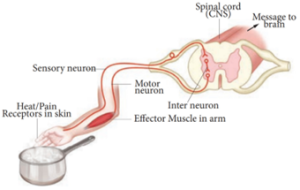
Reflex action and Reflex arc:
When dust falls in our eyes, the eyelids close immediately not waiting for our willingness; on touching a hot pan, the hand is withdrawn rapidly.
The spinal cord remains as a connecting functional nervous structure in between the brain and effector organs. But sometimes when a very quick response is needed, the spinal cord can effect motor initiation as the brain and brings about an effect. This rapid action by spinal cord is called reflex action. It is a fast, involuntary, unplanned sequence of actions that occurs in response to a particular stimulus. The nervous elements involved in carrying out the reflex action constitute a reflex arc or in other words the pathway followed by a nerve impulse to produce a reflex action is called a reflex arc.
Functional components of a reflex arc:
Sensory Receptor – It is a sensory structure that responds to a specific stimulus.
Sensory Neuron – This neuron takes the sensory impulse to the grey (afferent) matter of the spinal cord through the dorsal root of the spinal cord.
Interneurons – One or two interneurons may serve to transmit the impulses from the sensory neuron to the motor neuron.
Motor Neuron – it transmits impulse from CNS to the effector organ.
Effector Organs -It may be a muscle or gland which responds to the impulse received.
There are two types of reflexes. They are:
Unconditional reflex is an inborn reflex for an unconditioned stimulus. It does not need any past experience, knowledge or training to occur; Ex: blinking of an eye when a dust particle about to fall into it, sneezing and coughing due to foreign particle entering the nose or larynx.
Conditioned reflex is a response to a stimulus that has been acquired by learning. This does not naturally exist in animals. Only an experience makes it a part of the behavior. Example: excitement of salivary gland on seeing and smelling a food. The conditioned reflex was first demonstrated by the Russian physiologist Pavlov in his classical conditioning experiment in a dog. The cerebral cortex controls the conditioned reflex.
Peripheral Neural System (PNS):
PNS consists of all nervous tissue outside the CNS. Components of PNS include nerves, ganglia, enteric plexuses and sensory receptors. A nerve is a chord like structure that encloses several neurons inside. Ganglia (singular-ganglion) are small masses of nervous tissue, consisting primarily of neuron cell bodies and are located outside the brain and spinal cord. Enteric plexuses are extensive networks of neurons located in the walls of organs of the gastrointestinal tract. The neurons of these plexuses help in regulating the digestive system. The specialized structure that helps to respond to changes in the environment i.e. stimuli are called sensory receptor which triggers nerve impulses along the afferent fibres to CNS. PNS comprises of cranial nerves arising from the brain and spinal nerves arising from the spinal cord.
Cranial nerves: There are 12 pairs of cranial nerves, of which the first two pairs arise from the fore brain and the remaining 10 pairs from the mid brain. Other than the Vagus nerve, which extends into the abdomen, all cranial nerves serve the head and face.
Spinal nerves: 31 pairs of spinal nerves emerge out from the spinal cord through spaces called the intervertebral foramina found between the adjacent vertebrae. The spinal nerves are named according to the region of vertebral column from which they originate
- Cervical nerves (8 pairs)
- Thoracic nerves (12 pairs)
- Lumbar nerves (5 pairs)
- Sacral nerves (5 pairs)
- Coccygeal nerves (1 pair)
Each spinal nerve is a mixed nerve containing both afferent (sensory) and efferent (motor) fibres. It originates as two roots:1) a posterior dorsal root with a ganglion outside the spinal cord and 2) an anterior ventral root with no external ganglion.
Somatic neural system (SNS):
The somatic neural system (SNS or voluntary neural system) is the part of the peripheral neural system associated with the voluntary control of body movements via skeletal muscles. The sensory and motor nerves that innervate striated muscles form the somatic neural system. Major functions of the somatic neural system include voluntary movement of the muscles and organs, and reflex movements.
Autonomic Neural System:
The autonomic neural system is auto functioning and self governed. It is a part of peripheral neural system that innervates smooth muscles, glands and cardiac muscle. This system controls and coordinates the involuntary activities of various organs. ANS controlling centre is in the hypothalamus.